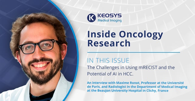
Maxime Ronot, Professor at the Université de Paris, and Radiologist in the Department of Medical Imaging at the Beaujon University Hospital in Clichy, France, has been working in Hepatocellular Carcinoma (HCC) since 2008. In 2021, he became a central reader for Keosys. We recently spoke with Prof. Ronot about the intricacies and challenges of using mRECIST to assess treatment response in HCC and his work as a Professor of Radiology. The following is a selection of excerpts from our conversation.
Keosys: What drew you to radiology and HCC?
MR: I knew I wanted to work in abdominal diseases, and the liver was appealing to me. Then it happened that when I was a med student, I came to this department. I liked the institution and the dynamics of the research, so I said, “Okay. This is what I want to do when I grow up.”
Keosys: And now you teach medical students how to read images and abide by the mRECIST criteria. How do you go about that?
MR: In France, it's case-based teaching. Residents read the exams first. Then a senior consultant, someone like me, reads the exam. The senior consultant will then correct the resident. Also, we have classical teaching situations where the residents will gather, and we will address specific points or answer specific questions.
Keosys: What are the challenges for these students?
MR: I think the most important challenge is to make them realize that they're not here to read images. They're here to treat patients. The main purpose of diagnostic radiologists is to use medical knowledge about the disease, about the course of evolution of the tumors, and the different kinds of treatments, to feed interpretations and make them richer and more focused on the point.
Keosys: Given this, how do you approach an image in the context of reading for a clinical trial? After all, when you're performing central reads, you don't have clinical data.
MR: In that case, we focus mainly on the pure assessment of response measurements, target selection, etc. And when I do that, I really want to make sure that there is, for instance, a progression when there is a progression. That's what I think about when I do central reads.
Keosys: What are the challenges in using the mRECIST criteria?
MR: There is a problem in that those criteria have not been deduced by observation and scientific reasoning. They've been proposed based on previous criteria that have been more widely validated. So they introduced a new concept—imaging-based viability of a tumor corresponding to the enhancement of the tumor on arterial phase. But then they said, “Okay, let's just use the same thresholds, the same rules, and everything is going to be the same except for the viable part of the tumor.” That’s not really scientific. Now, 12 years after the introduction of those criteria, we've seen that we have a few problems. First, what should we do when the tumor does not show an enhancement on arterial phase? I know that the tumor is viable, I know that this is not a dead cancer, but there is no enhancement on arterial phase. So technically, I cannot use the mRECIST criteria. I can still assess tumor response, by switching to RECIST.
Keosys: So where do we go from here?
MR: That's a difficult question. Our problem is that we see there is more and more discrepancy between the way the tumor behaves and the outcome of patients, especially when you use immunotherapy. You can have stable disease for months and months, or some patients will progress, but you stick with the treatment and in the end, it will benefit the patients. I am not sure that iRECIST, the criteria that are designed for immunotherapy, are going to be really useful in HCC, because the occurrence of pseudo progression seems to be extremely rare. So, probably the benefit of those criteria is not needed in HCC patients. Maybe that's a possible simplification in the years to come. But we will keep the mRECIST part, I guess, because we still use molecules that target vascularization of tumors. So, I think this one is going to stay.
Keosys: There is a lot of conversation around the potential impact of artificial intelligence (AI) on the reading of images. Any comment on that?
MR: Is AI going to help us to assess response? I think so. There are interesting initiatives underway to try to help radiologists detect the lesions and registered images from one exam to the next and things like that, or summarize, and then help us or suggest, and I know there are companies that already have proposed some semi-automated, AI-based solutions.
Keosys: Would you be using AI to measure diameters or volumes? Or to detect viability in a way?
MR: When we assess the response, we assume that the amount of tumor is directly related to the amount of life. So, monitoring the changes in tumor burden is a way for us to know, more or less, if the patient is going to survive. That's an assumption that we make. What we would like to do is monitor everything and not be forced to choose just a couple targets. Right now, we say, “Okay, tumor burden is too complicated. So, let's do a selection of tumor burden. And measuring the volume is too complicated, so let's do a measurement of size. And then two dimensions is too complicated, so let's do measurement of one.” We’ve created a cascade of assumptions and gotten further and further away from the original one. Maybe AI will help us go back to what we initially wanted to do: Quantify the entire tumor burden. If we do that, we can assume that our predictions will be more accurate. So maybe that's one of the things AI will help us do, to get rid of all the assumptions that we are forced to make along the way and get closer to the initial assumption, that tumor burden means life.


