Neuroendocrine tumors (NETs) arise from hormone-producing cells that form part of the neuroendocrine system. Neuroendocrine cells have dual functions: they act as hormone-producing cells and nerve cells. Their role is to receive signals from the nervous system and respond by releasing hormones to perform specific functions. For instance, neuroendocrine cells control how fast food moves through the gut, in the lungs they regulate air and blood flow, and in the pancreas they control insulin release to lower glucose in blood.

Given that the neuroendocrine system is distributed throughout the body, NETs can be scattered anywhere in the body. Most, however, originate in one of four sites:
- Appendix
- Small intestine
- Rectum
- Lungs
Because an endocrine tumor develops from cells that produce hormones it can, secrete excessive amounts of hormones, which may result in serious illness.
NETs are relatively rare: even taking the different sites of origin into account, they make up ~2% of all malignancies. The annual NET incidence is 6.98 per 100,000 persons, and appears to be rising, probably due to progress in early detection.
Two Tier Diagnosis
Since their discovery at the beginning of last century, the debate over how to diagnose and grade NETs has been raging. But a consensus seems to be growing that NETs fall into two broad categories:
- Low-grade, well-differentiated tumors. Probably benign.
- High-grade, poorly differentiated, rapidly proliferating, aggressive carcinomas. Poor prognosis.
NETs are further classified in grades, following SEER histologic: G1, well-differentiated, G2, moderately differentiated, G3, poorly differentiated and G4 undifferentiated or anaplastic. It’s worth noting that some NETs defy their histological classification, and although grade 1 or 2, that predicts a low-risk, slow evolution, they metastasize and transform like a grade 3 tumor.
To Localize a NET
Imaging is critical because, if a tumor is caught early, surgery can be curative for non-metastatic disease.
The techniques of choice are computed tomography (CT) and endoscopy. Other imaging methods include magnetic resonance imaging (MRI), ultrasonography, scintigraphy, and positron emission tomography (PET).
Which to select may depend on the anatomical site:
- Bronchial NETs: a chest radiography followed by CT or MRI. A bronchoscopy has the advantage of providing a biopsy
- Rectal, duodenal, colonic and gastric tumors: endoscopy is the preferred method. But at the early stages, when tumors are small and difficult to detect, angiography can be used because NETs are hyper-vascularized.
- Gastro-entero-pancreatic NETs, which are mostly asymptomatic and often discovered during surgery for another reason, may need scintigraphy and a somatostatin analog for visualization. [See ‘Find and treat’ below]
- Metastases require MRI, CT scans or ultrasonography.
Find and Treat: Radiolabelled Agents
NETs are particularly amenable to imaging with radioactive tracers because the tumors overexpress certain markers. For instance, somatostatin receptors are highly expressed on the majority of NET cells. A technique called Octreoscan uses the synthetic somatostatin analogues, octreotide or lanreotide, labelled with Indium-111. When injected into the patient, the radioactive analogues bind the somatostatin receptors and allow their visualization. For visualizing pheochromocytomas, a rare type of adrenal gland NET, an Iodine-123 labelled noradrenalin analogue is the best suited to the job.
Although scintigraphy cannot approximate tumor size, it can point to the extent of metastasis and indicate tumor location.
Another tool to detect early stage NETs is Netspot, marketed by AAA (a Novartis company) as a companion diagnostic to Lutathera (See ‘What treatments are available for NETs’ below). This PET diagnostic kit combines, a somatostatin analogue bound to a Gallium-68 with a covalently bonded chelator DOTA-TATE,
Ipsen is also developing a Gallium-68 satoreotide (OPS202) for monitoring and diagnosing gastro-entero-pancreatic-NETs using PET imaging. The corresponding phase II trial is currently recruiting participants. And RadioMedix and partner Curium are recruiting patients for a phase III trial to evaluate Copper- 64-Dotatate as a PET-CT diagnostic agent.
What Treatments Are Available for NETs?
Most NETs are treated with surgery, especially low to intermediate grade tumors. If, however, surgery is not feasible because metastases have developed, systemic options are available.
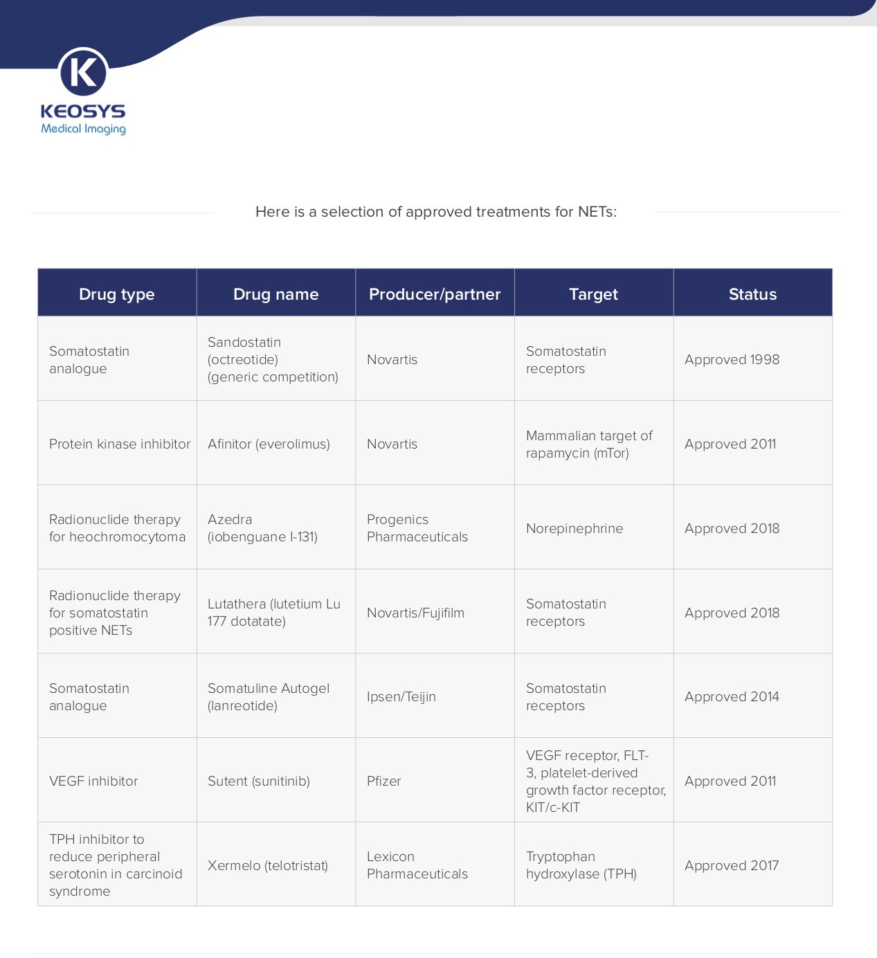
The table below highlights a selection of approved treatments for NETs. (Click here for an enlarged version.)
The next stage in clinical trials is to test combinations for synergistic effects, for example octreotide and bevacizumab (Avastin), a cancer treatment that targets vascular endothelial growth factor A (VEGF-A). Clinical trials with sunitinib and everolimus are underway. Also, an immunomodulatory checkpoint inhibitor from Novartis that targets programmed death-1 receptor (PD-1) is in phase II testing.
An investigative next-generation radiolabelled peptide receptor therapy developed by Ipsen, OPS201 (Satoreotide) labelled with Lutentium 177 is in clinical testing for metastatic somatostatin positive NETs.
Keosys' Role in the NET Clinical Trials
Keosys is involved in managing several early and late phase clinical trials for NETs, and played a role in a recent FDA and EMA approval of a radioactively labelled somatostatin analogue that not only enables visualization and diagnosis, but also allows treatment in patients with inoperable progressive somatostatin receptor positive tumors.
For these trials, Keosys manages the Blinded Central Independent Reading processto provide results characterizing the molecules being studied. (see further information in our December 12, 2018 blog: “Recent Drug Approvals Based on Imaging Results”).
Our IMAGYS medical imaging software supports the study-specific patient eligibility confirmation and the efficacy analyses ensuring a smooth collaboration between radiologists and nuclear medicine physicians.. The system also enables medical images’ centralization and quality control.
Keosys technology and expertise is also successfully applied to manage dosimetry sub-studies during these trials (see further information in our January 22, 2019 blog, “Dosimetry and Its Use in Clinical Trials”).
The Future for NETs
Radiopharmaceuticals are emerging as the next frontier in NET treatment, not just as diagnostic agents but also as therapeutics. Two such agents have recently been approved: Lutathera and Azedra (see Table).
Combining such radionuclide therapies with molecular imaging allows dosing to be customized to each patient, a strategy that may achieve the ultimate challenge of irradiating even small tumors with little or no damage to surrounding healthy tissues.